Thursday, November 9, 2017

Winning the Robert Narins Award

The Award


Saturday November 4, 2017 I accepted the Robert Narins Award from the ASN. This is the highest honor I could ever receive for the work that I have done in medical education.

Here is how ASN describes the award:
The Robert G. Narins Award honors individuals who have made substantial and meritorious contributions in education and teaching. This award is named for Robert G. Narins, who is also the first recipient of the award. 
Dr. Narins' contributions to education and teaching started in 1967 when he was appointed to the faculty of the University of Pennsylvania. At Penn, and on the faculties of UCLA, Harvard, Temple and Henry Ford Hospital, he taught and mentored many residents and fellows. For eight years he chaired the ABIM's Nephrology Board and also worked on the ACP's Annual Program Committee. His contributions to education in the fields of fluid-electrolyte and acid-base physiology are prodigious and well-recognized. 
Dr. Narins was also involved in the creation and planning of many ASN educational programs during Renal Week and throughout the year, including: Board Review Course and Update, one and two day programs at Renal Week, Renal WeekEnds, and NephSAP. He also was instrumental in the decision to develop the Clinical Journal of the American Society of Nephrology (CJASN), the establishment of the Fellow of the American Society of Nephrology (FASN) program, and negotiated the successful partnership agreements with HDCN and UpToDate. Dr. Narins has been at the forefront of collaborative efforts with the American College of Physicians to increase the exposure of nephrologists to relevant updates in Internal Medicine and internists to chronic kidney disease. Collaborative educational programs with societies in Europe and Asia have helped to spread education and teaching in nephrology on a global scale.
To be recognized for this is amazing. I feel that social media and internet-based, distributed learning that leverages the skills and perspective of a large group of self-appointed experts is, and will, continue to swamp the old model of a few, highly selected, experts.

The future of medical education will not be televised. It will be streamed

In many ways, the reality of internet distributed medical education is the opposite of what people believed it would represent. I remember hearing people speak about the best lecturers being able to distribute their wisdom to the world through IP switching. The conventional wisdom was that the internet would allow all medical students to learn from the best teachers. The internet would eliminate location and size limits of the lecture hall so we would all get the Harvard education.

Social media altered that future. 

Instead of one vision being distributed to a million impressionable minds we got a million visions being distributed so that each mind could find the one that worked for them.

We are in the midst of a massive democratization of medical media.

Social media in medical education is moving us from broadcast video to YouTube, from radio and audio books to podcasts, from books and journals to blogs and tweets, from a few highly selected authority figures to a large number of educator hobbyists. 

And I am just a participant in this. I am an early adopter. I am at the front of the wave. However, I didn't create the wave, and I can't control it; I'm merely surfing the leading edge. And I believe ASN recognizes that. I believe that the ASN awarded me the Narins award as a symbol that distributed, social media-powered-learning, is now a valid and growing part of primary and continuing medical education. ASN is among the first of the medical societies to recognize this. I salute ASN for resisting the urge to circle the wagons and fight the inevitable. ASN should be commended for recognizing and embracing this new epoch in medical education.

That said, as much as I believe I was a just a symbol of the revolution that I am part of, they did pick me to be that symbol and for that I am grateful. And I would like to thank the people that have brought me on this journey. 


That tweet is totally true. And in the rush to compose a coherent thank-you speech in my adrenalin addled brain I glossed over and missed some essential people.

The Patron Saint

Burton D. Rose. My favorite part of the Narins Award Legacy Video is when Rose says that Robert Narins brought fluid and electrolyte and acid-base teaching into the mainstream (1:35) and set the path for him.


I love this because Rose set the path for me, in two ways. 

One: Rose's Clinical Physiology of Acid-Base and Electrolyte Disorders was the bible that taught me renal physiology and inspired me to pursue a career in nephrology. I bought it during my third year of medical school when I told the intern that I was working with that I was confused by eletrolytes. He suggested this book. Talk about a bad answer to a small question. I wanted a 5-minute lecture on what fluid to order and my senior suggested a 916 page, $70 tome.

But I was an idiot and I bought it.

And though it sat on my shelf for a year before I started it, when I began, I could barely put it down. I truly believe that one can draw a direct line from that terrible advice given to me at the Allen Park VA in 1993 to my position today. Sometimes bad advice can lead to the greatest of outcomes.

Rose's book is a masterpiece because it strives to make the reader build a robust mental model of how the kidney works. Once that model is complete, it becomes easy to understand all of the electrolyte disorders.

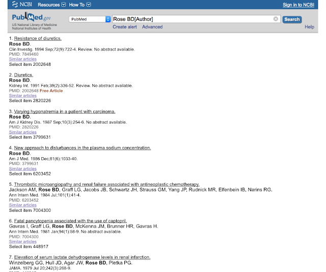
Two: When I was an intern I was telling a doctor how much I loved Rose's yellow electrolyte book and he told me to look him up in PubMed. I had previously looked up Berl and Schrier and I expected much of the same, but this is what I got:


Seven. Seven articles in pubmed by the great Bud Rose. The man that has done more for medical education and patient care than anybody since Osler (based on his work with UpToDate) had seven articles in PubMed in 1996. Go ahead, see for yourself. This convinced me that I could be a medical educator without being a medical researcher. I could pursue what I loved, without being distracted doing what I had little interest in doing.

The Inspiration

Joshua Schwimmer. Josh was the first nephrology blogger. I had been a long time reader for years before following his trail and starting Precious Bodily Fluids. And after a few months, Joshua gave me my first link and turned the microphone on. Thanks Josh.

The Collaborators


PBFluids and @Kidney_Boy stand apart as the only signifigant projects I have done alone. Everything else of significance has been done with a collaborator, co-author, or co-creator. This is an indisputable case of "I couldn't have done it alone."

Sarah Faubel as a co-author on the Microbiology Companion and The Fluid Electrolyte and Acid Base Companion

Joel Smith as co-creator of Alert and Oriented Publishing

Burke Mamlin for the work on Kidometer

Kenar Jhaveri for choosing me to be part of AJKDblog

Matt Sparks for NephMadness, DreamRCT, NSMC

Swapnil Hiremath for NephJC, NSMC

Edgar Lerma for Nephrology Secrets

Anna Burgner and Tim Yau for NephMadness

The Crew


#NephTwitter is a colorful and engaging place with an always on conversation anchored by people around the world. Here are a few of the people that populate that community.

Tom Oates, Roger Rodby, Paul Phelan, Michelle Rheault, Francesco Iannuzzella, Hector Madariaga, Graham Abra, Nikhil Shah, Matt Graham-Brown, Ian Logan, Scherly Leon, Dearbhla Kelly, Silvi Shah, David Goldfarb, Richard McCrory, F. Perry Wilson, Raymond Hsu, Benjamin Stewart, Brian Stotter.

The Enablers


My partners at St Clair Specialty have been involuntary partners on this journey and have been great. St Clair Specialty is a practice that values people that contribute to nephrology. This was clear from past CEO, Robert Provenzano, who was a major contributor to nephrology as a scientist and president of the RPA. And it is also clear with Keith Bellovich, RPA board member and chief of nephrology at St John Hospital and Medical Center. At a more single minded-practice, I could not have contributed to nephrology education as I have.

And lastly, and most importantly, I could never have contributed to medical education without my family. I need to thank the eternal patience and understanding of my wife, Cathy, and my kids, Laura and Simon Topf. Thank-you.



Friday, October 27, 2017

Site improvement

If you have been annoyed at all of the broken links at PBFluids. My apologies. Blogger doesn't host any files so to use them you need to host elsewhere. For this reason alone I recommend WordPress or SquareSpace for people who want to start blogging.

Tonight I fixed the Handouts tab. A few weeks ago I fixed the Books tab. All of those links now work. Next up: the Lectures tab.

Question: Should old lectures that may contain out of date material be taken down, or remain up?

Tuesday, October 24, 2017

Fuck you Wikipedia

The Wikipedia entry for Journal Club used to have a comprehensive list of Twitter-based Journal Clubs. It looked like this


I went to add another journal club to the list yesterday (hello Journal of Hospital Medicine and #JHMChat) and it was gone. I went into the history and discovered that Twitter Journal Clubs are not notable so on September 28th that list was taken from the world. Sorry. 

I'm thinking of a new Wikipedia tag line, how does this sound: Wikipedia, where a comprehensive list of porographic actresses belongs in the encyclopedia, but Twitter Journal Clubs? Not notable.


Monday, October 23, 2017

Using visual abstracts in presentations

This past Saturday I gave a talk at the ACP of Michigan on SGLT2 inhibitors.

The talk went well, except my HDMI to thunderbolt converter failed in a big way and I had to export the presentation to Powerpoint and run it off a Windows Machine. Yuck.


I used visual abstracts from EMPA-REG and CANVAS as a significant part of the presentation. This segment demonstrates how I used them. What killed me was the cool animation, where the third panel flips to reveal the renal outcomes, was handled with complete incompetence by PowerPoint. Otherwise PowerPoint did a pretty good job displaying my slide, but botching my favorite animation in the entire presentation is bordering on unforgivable.


SGLT2i renal outcomes from joel topf on Vimeo.

Here are the two visual abstracts in question:


This slide isn't in the above video. I will eventually get the whole presentation up, but I love this one so much I had to share.


Speaking of sharing. This Wednesday, Dr. Christos Argyropoulos will kick off the first Tubular Talk with a presentation on SGLT2 inhibitors. Should be great check out all of the details at GlomCon.


Monday, October 9, 2017

Things I want to write about eventually: Exercise induced rhabdomyolysis

Exertional Rhabdomyolysis during a 246-km Continuous Running Race

SKENDERI, K. P., S. A. KAVOURAS, C. A. ANASTASIOU, N. YIANNAKOURIS and A. MATALAS. Exertional Rhabdomyolysis during a 246-km Continuous Running Race. Med. Sci. Sports Exerc., Vol. 38, No. 6, pp. 1054 – 1057, 2006. Background: To evaluate the effect of continuous, moderate-intensity ultraendurance running exercise on skeletal muscle and hepatic damage, as indicated by serum enzyme activity measured immediately following the race. Methods: Thirty-nine runners of the Spartathlon race (a 246-km continuous race from Athens to Sparta, Greece) who managed to complete the race within the 36-h limit participated in this study. Mean finishing time of the study participants was 33.3 T 0.5 h and their average age, height, and body mass were 41 T 1 yr, 174 T 1 cm, and 67.5 T 1.1 kg, respectively. Blood samples, taken a day before and immediately after completion of the race, were assayed for the following variables: creatine kinase (CK), lactate dehydrogenase (LDH), aspartate aminotransferase (AST), alanine aminotransferase (ALT), and gamma-glutamyltransferase (F-GT). Results: A dramatic increase in most of muscle and liver damage indicators was observed. The mean values for CK, LDH, AST, and ALT after the race were 43,763 T 6,764, 2,300 T 285, 1,182 T 165, and 264 T 37 IUILj1, respectively. These values were 29,384 T 4,327, 585 T 89, 5,615 T 902, and 1,606 T 331% higher than the corresponding values before the race (P G 0.001) for CK, LDH, AST, and ALT, respectively. However, there was not a significant increase in F-GT levels. Conclusion: Muscle and liver damage indicators were elevated at the highest level ever reported as a result of prolonged exercise, although no severe symptoms that required hospitalization were observed in any of the participants. The data suggest that even moderate-intensity exercise of prolonged duration can induce asymptomatic exertional rhabdomyolysis. Key Words: CREATINE KINASE, LACTATE DEHYDROGENASE, SPARTATHLON, ULTRAENDURANCE EXERCISE

So tasty.


Link


Thursday, October 5, 2017

Get the NephRUN T-shirt While Fighting Multiple Myeloma

This tweet has taken off.


A few people have expressed an interest in getting one for themselves. Okay, I'm a reasonable guy. How about this deal.

If you are going to Kidney Week and you donate $50 to the Multiple Myeloma Research Foundation for my trip to Everest I will get you a t-shirt. Your donation is even tax-deductible. This needs to happen in the next 10 days for me to get the shirts in time.

If you are not going to Kidney Week, donate $100 and I will send you a shirt.

Want a shirt and you already donated to the MMRF? Shoot me a tweet (DM or @) or e-mail and we'll work this out.

After you have donated, fill out this form so I get you the right shirt and know how to get it delivered.

The shirt is really nice. The women's version is a Hanes Ladies Cool Dri V-Neck Performance Shirt and the mens is the same shirt, but crew neck.

Thursday, September 28, 2017

Have a nephrology question? #AskRenal to the rescue.

A couple of days ago, this came across my notifications


The answer came quick.


The video is here (complete with misspellings) and the Keynote file is here (with misspellings corrected)


How metabolic alkalosis causes hypokalemia from joel topf on Vimeo.

Monday, September 25, 2017

The beautiful #VisualAbstracts of the NEJM

This summer (I think) the NEJM began publishing visual abstracts on their twitter feed. Curiously, I was unable to find them on the page of the article that the visual abstracts references, or in the list of media types that you can search for.

The figure list on the right side does not include the striking visual abstract they created.


The "Browse Figures and Multimedia page has 19 different types of media, but visual abstract is not one of them.

The only way I could round up the visual abstracts was scrolling through the the NEJM Twitter feed. Here are the ones I found. Did I miss any?


Gorgeous work. Each one has a unique color palette and they have a pretty simple template, but three different ways of executing it. All of them look like they are from the same family except the tiotropium visual abstract. I really like that they give both the percentages and the raw numbers. No P-values or confidence intervals are found. These visual abstracts have as low an information density as I have seen. This is not a criticism, I think meh style has been increasing complexity to the detriment of my work. I need to turn up my inner NEJM.


Monday, September 11, 2017

Another question from OUWB

Hi Dr. Topf 
First of all, apologies for sending this via email but I do not have a Twitter account (I know, its the 21st century, who doesn't?). 
I had a quick question regarding a practice problem I was doing. Rather than summarize the question for you, I included a screenshot so that you have the primary source with the explanation provided. Below, I also included my explanation for my reasoning for choosing that option. Basically, I am confused as to why the bicarb would be decreased in this scenario.


So the stem describes acute trauma. Specifically crush injuries, so you should be thinking rhabdomyolysis where the body gets turned inside out. In my very first lecture we talked about the intracellular atmosphere versus the extracellular atmosphere:


So expect increased potassium and phosphorus.

The vital signs show a patient with circulatory insufficiency, i.e. shock. There are some initial labs drawn from the blood and urine right before resuscitation is initiated. The urine shows an osmolality of 800 mmol/kg H2O (highly concentrated, indicating a lot of ADH activity) and a urine sodium of 5 mEq/L (very low, indicating increased activity of the renin angiotensin aldosterone system). 

The question then asks you to predict the serum labs. Cool question. The best testing strategy here is to cross off ones that make no sense. Here are the foils:


BUN. This should be elevated as the patient moves to a pre-renal physiology. This leads to an increased filtration fraction to maintain GFR in the face of decreased renal plasma flow. This causes an increase in the osmolality in the efferent arteriole and vasa recta. this increases osmotic reabsorption of fluid from the proximal tubule. BUN flows passively with the fluid, decreasing renal urea clearance and increasing serum BUN. So D is wrong. E is wrong.

Potassium (K+) ions. All the choices show that it rises as the rhabdomyolysis from the crush injury releases loads of intracellular potassium. No answers are eliminated here.

Sodium (Na+) ions. We have a mishmash of choices here. This is difficult to predict. The increased ADH released due to shock would tend to lower the sodium. Increased aldosterone and renin would tend to increase the sodium. Since both are happening together I would expect them to balance each other out resulting in no change in sodium concentration. This is especially true since the stem specifically says there has not been much urine output I would go with D, for no change in sodium concentration. NOTE: About activation of the RAAS as a cause of hypernatremia. Hypernatremia is commonly listed as a symptom of Cushing syndrome and hyperaldosteronism so it is possible to have hypernatremia from (at least the pathological) activation of the renin angiotensin aldosterone system) but this is very unusual as increased in sodium concentration stimulates thirst which dilutes the sodium back to normal. 

THIS COLUMN IS MESSED UP. THIS IS AN ERROR BY THE QUESTION AUTHOR.

Hydrogen (H+) ions. All the choices show that it rises. In shock we expect an increase in hydrogen ions as patients move to anaerobic metabolism due to inadequate perfusion (the functional definition of shock). No answers are eliminated here.

Bicarbonate (HCO3-) ions. Bicarbonate is the primary buffer in the body. Increases in hydrogen ions will be buffered by bicarbonate and bicarbonate will be consumed.
So the right answer is bicarbonate will be decreased. This eliminates answer A.

This leaves us with B or C and the question hangs on what the sodium will do and the reality is the change in sodium is unknowable. Shit question. Sorry.

Here is what Kaplan claims to be true:

The forth bullet point is the one where the question fails. It is true that there is accumulation of plasma electrolytes in acute kidney injury. The problem is that these are electrolytes are measured as concentrations and there is also an accumulation of water (which is normally excreted by the kidney). This means that the effect on concentration is variable. Some, like hydrogena and potassium, reliably increase in AKI, but sodium is often decreased in AKI. Maybe the question writers were looking at an unconventional way measuring ions in the the plasma (as total amount rather than concentration).

The line in the answer that pre-renal azotemia is associated with hypernatremia is just wrong. You will encounter numerous patients with pre-renal disease that will have simultaneous hyponatremia. It is impossible to predict the serum sodium concentration from the volume status. This question reinforces the worst instincts of med students when it comes to predicting serum sodium. As I emphasized in the lecture, volume regulation and osmoregulation have two different regulatory systems and, though there is some cross talk, they are largely independent from each other.

The last paragraph tries to make the case that hyponatremia is more common in ATN while hypernatremia predominates in pre-azotemia. This is total fiction and does not exist. Though there can be more urinary sodium in ATN, if the patient is oliguric, it doesn't matter how high the urine sodium concentration is, if the urine volume volume is close to zero there will not be much urinary sodium excretion.

Distinguishing between pre-renal azotemia and ATN is a constant problem in nephrology. Trust me you can't make the determination by looking at the serum sodium. It aint that easy.

This question writer should never write another question about sodium. 

 I posted this to twitter. The subsequent discussion was pretty interesting:


Thursday, September 7, 2017

Medical student questions about nephrology

I have the honor of teaching at Oakland University William Beaumont School of Medicine. I teach sodium and water and acid-base to the second year medical students. After the lectures there is a steady stream of questions that start to fill my in-box. I answer the e-mail but I also post the questions and answers on PBFluids. Here is a directory to this year's crop of Q&A.

Sodium concentration versus sodium content. With a second question on pseudohyponatremia vs false hyponatremia vs factitious hyponatremia

Macula densa and TG feedback. With a second question on whether SIADH is really euvolemic or just mostly euvolemic.

Breaking down an acid-base question.

More on euvolemic hyponatremia and how does this affect uric acid.

Urine chloride in non-anion gap metabolic acidosis, where does it come from?

Starling forces and GFR. With additional Q&A on edema, and metabolic acidosis and ammonia-genesis.


Wednesday, September 6, 2017

The Fluid Electrolyte and Acid Base Companion

After Medical School Sarah Faubel and I set out to explain sodium, potassium and acid-base in a programmed text. Four years later, in late 1999 we finished this journey. The result was The Fluid, Electrolyte and Acid Base Companion.

5 stars on Amazon


Highly reviewed by Beaver Medic


Also by the Mark Yoffe.
So where do you get this book? Right here, for free (PDF, 29 mb):


If you would prefer a zip file with each chapter in individual PDF files, click here.

Other links about The Companion:

Dropbox problems

A while ago Dropbox changed how they handled public links. Then they announced that old links with the previous public folder system would be unsupported. I have no idea how many or where these links are littered through out PBFluids, but I suspect as of September 1, there will be a lot of them.


Today I received this tweet.


So this bug struck the prized link on the whole damn blog. Annoying. Blogger is the only platform that doesn't host files and forces you to store them elsewhere. This is the root cause of the problem. WordPress and SquareSpace allow you to host your files on their servers so you wouldn't run into this problem reconnecting two services.

That's like strike seven against Blogger. I've got to get out of this burning pile.

Monday, August 28, 2017

OUWB Starling forces question


I was hoping I could ask you a few questions. I’m finding there is a lottttt of contradictory information.
  1. According to starling forces, decreased plasma oncotic pressure should increase GFR, but according to nephrotic syndrome, decreased albumin will cause edema and overall decrease GFR. Which one should I believe? 
  2. In general, it’s said that AT2 at low levels dilates the afferent arteriole to increase GFR, but at high level it constricts both efferent and afferent to decrease GFR. However, the SNS, which stimulates renin, constricts all arterioles in the body as well as activates the RAAS system. How does that work? Is the SNS more immediate until the aldosterone system is ready to say okay go ahead and dilate the afferent I’m ready to take up the water anyway? 
  3. This is a very basic question but sometimes I have moments of self doubt and this is one of them: So we always say edema is fluid buildup in ISF due to increased hydrostatic or decreased oncotic pressure (like nephritic syndrome hypoalbumineia) right? So why does fluid build up in ISF as opposed to go inside the cell where I guess technically there is more stuff to pull it in? 
  4. How does K suppress ammonia genesis? 

Thank you very much!


Let's take these one by one,

NUMBER ONE
Nephrotic syndrome and GFR. Don't connect those neurons. Proteinuria does not cause an immediate and hemodynamic change in GFR that is clinically meaningful. Yes, you are right that lower oncotic pressure should increase GFR, but those increases in GFR will be trimmed by tubuloglomerular feedback so that in the end there is not a meaningful change in GFR. Likewise the nephrotic syndrome will cause fluid to leak from the blood vessels decreasing effective circulating volume lowering renal plasma flow. However, once again these changes in volume are small enough that the kidney easily compensates with changes in AT2, PGE, filtration fraction, etc so that GFR remains stable.

Over a long time, proteinuria causes chronic kidney disease and decreases renal function, but not by the mechanisms you described.

Of note the model you are talking about with nephrotic syndrome causing fluid to leave the blood vessels and that resulting in decreased perfusion of the kidney is a model called underfill hypothesis of edema in nephrotic syndrome. Most nephrologists now ascribe by the overfill hypothesis which states that the primary abnormality is not loss of fluid from the capillaries from the decreased albumin, but increased sodium absorption by the diseased kidney. This results in volume overload and that causes the edema.

NUMBER TWO

As I understand it angiotensin is only a vasoconstrictor. The proximal tubule is dilated by prostaglandin E. In volume depletion there is release of renin which activates angiotensin 2 (with help of angiotensin converting enzyme). Angiotensin 2 vasoconstricts both the afferent arteriole and efferent arteriole. But since the afferent arteriole is so much bigger to begin with, after the angiotensin 2 induced vasoconstriction the resistance in the afferent arteriole is less than the resistance in the efferent arteriole, this serves to increase the intraglomerular pressure, forcing more plasma through the glomerular slit membranes and increasing the filtration fraction and maintaining GFR in the face of volume depletion.

And yes the SNS is more immediate and the renin angiotensin aldosterone system is a bit slower.

NUMBER THREE

Where fluid builds up depends on what is being altered. In nephrotic syndrome, the (underfill) theory states (I'm an overfill believer) that decreased plasma albumin lowers the oncotic pressure drawing fluid from the interstium back into the capillaries at the venous end. This means more of the fluid remains in the interstium leading to edema. The oncotic agent of note here is albumin which determines the flux of fluid between the interstitial and plasma compartment.

In order to shift fluid between the intracellular and extracellular compartments you would need to change sodium and potassium which are the chief osmotically active particles of interest between those two compartments.

NUMBER FOUR

Hyperkalemia causes potassium to shift into the cells. To maintain electroneutrality hydrogen leaves the cell. One cation in, one cation out. The loss of hydrogen ions makes the cell alkalotic. This rise in pH tricks the proximal tubule cell into believing the entire body is suffering from metabolic alkalosis and since ammonia generation is used to increase hydrogen excretion, and correct metabolic acidosis, metabolic alkalosis shuts down ammonia generation.

Sunday, August 27, 2017

OUWB Non-anion gap question

Where does the chloride come from.

XXXXX and I had a question following the Acid-Base workshop. What is the origin of the increase in chloride ions in patients with NAGMA due to GI or renal causes?  
Thanks, 

So the key here is not to think of the body as static. As patients lose bicarb in the stool or in the urine, this will result in volume depletion which will be compensated for by renal retention of sodium and yes, chloride.

Great review of non-anion gap metabolic acidosis here:

OUWB Euvolemic Hyponatremia question

Hello Dr. Topf, 
I hope you are enjoying your weekend. I had a question in regards to one of your lectures. I was wondering why there is a low level of Uris acid in euvolemic hyponatremia but not in hypervolemic or hypovolemic hyponatremia. Also, how is it that Na taken in equals Na excreted in euvolemic hyponatremia? 
All the best,


So why is there a low level of uric acid with euvolemic hyponatremia? Let's first look at what happens to uric acid in the other causes of hyponatremia, namely hypovolemic and hypervolemic. In both of these situations the kidney is experiencing decreased perfusion, either from absolute volume depletion (diuretics, diarrhea) or perceived volume depletion from pump failure (CHF) or fluid maldistribution (cirrhosis and nephrotic syndrome). 

In these volume depleted states there is an increase in the filtration fraction, i.e. more of the plasma that enters the glomerulus is actually filtered. This is how the kidney compensates for a decrease in renal plasma flow while maintaining GFR, it increases the fraction of fluid that is filtered. 


A consequence of this, is that the oncotic pressure in the blood leaving the glomerulus is higher because more of the fluid (but none of the protein) has gone down the glomerular drain leaving the plasma in the efferent arterioles with a higher oncotic pressure. 

This plasma then enters the vasa recta where it surrounds on the proximal tubule. Here the increased oncotic pressure pulls more fluid back. 

This is an ideal situation. The increased filtration fraction maintains GFR in the face of decreased renal plasma flow, and the increased filtration fraction results in enhanced reabsorption of fluid in the proximal tubule limiting fluid loss in situations where patients have decreased perfusion.


Uric acid handling is complex and not fully worked out. 

It appears that there is both uric acid secretion and reabsorption in the proximal tubule. 

Functionally, uric acid clearance tracks with renal perfusion:
  • Decreased uric acid clearance with decreased renal perfusion
  • Increased uric acid clearance with increased perfusion of the kidney
This is similar to what we see with urea. The following description of urea handling gives a model that will work for uric acid, though the truth of uric handling is much more complex.

The key with urea is that it's handling in the proximal tubule tracks with total fluid reabsorption in the proximal tubule. 

With volume depletion, increased filtration fraction causes increased oncotic pressure in the vasa-recta increasing urea reabsorption in the proximal tubule.

In volume overload, decreased angiotensin 2 decreases sodium reabsorption resulting in less fluid reabsorption and less passive reabsorption of urea  so increased urea loss in the urine and lower serum urea.

Now what happens in euvolemic hyponatremia.

Sodium in equals sodium out. This means that these patients do not have a primary volume abnormality as we see in the hypovolemic and hypervolemic patients. Because of this their sodium regulation volume regulation system is not stressed, they are at homeostasis with regards to body sodium. When you are in homeostasis, in order to stay in homeostasis you need to excrete all the sodium that comes in. In other words sodium in equals sodium out.

However these patients are not in water balance. they have a disease that forces their ADH to 11. They have a fixed ADH secretion and it is set at full blast. This minimizes urinary water excretion, but they are able to stay in sodium balance. So the net of this is they make only a little bit of urine but that small amount of urine carries all of their ingested sodium (sodium in = sodium out) so the sodium is excreted in a small volume at a high concentration.

Now the obvious problem here is that they are holding on to an excess of water. And that will increase their total body volume. This is subtle and doesn't cause edema, or heart failure, or fluid overload in the lungs, but it is there. This fluid overload suppresses angiotensin 2 and decrease sodium resorption in the proximal tubule and hence decreases urea (and uric acid in our model) reabsorption. 

And yes this does mean it is not exactly sodium in = sodium out, there will be a slight excess of sodium excretion. 

OUWB Question: Acid-Base

Hi Dr. Topf,
(I don't have Twitter) I wanted to ask you about question 6 on the week 2 quiz:
"An unresponsive woman is brought to the emergency room. She has a history of a suicide attempt a few years earlier. The lab tests are: Serum Na 140 mmol/L Serum K 4.0 mmol/L Serum Cl 100 mmol/L Serum HCO3 14 mmol/L, BUN 17 mg/dl, creatinine 0.7 mg/dL, serum osmolality 323 mOsm/Kg, Blood glucose 72 mg/dl, Blood gases: pH 7.28 pCO2 27 mmHg. What would you expect the urine pH to be in this patient?"
Why is it that we would expect the urine pH to be acidic? Since blood pH is 7.28, I would imagine that urinating out HCO3- (explaining the low serum HCO3) would have caused the acidic blood pH, thus making urine pH basic?

Thanks for your help,


When answering multiple choice board-style question try to figure out what they are looking for. Let's break this down.

"An unresponsive woman is brought to the emergency room. She has a history of a suicide attempt a few years earlier. 

This is the “tell” of the stem. Acid base + suicide = ethylene glycol toxicity
The lab tests are: Serum Na 140 mmol/L Serum K 4.0 mmol/L Serum Cl 100 mmol/L Serum HCO3 14 mmol/L, BUN 17 mg/dl, creatinine 0.7 mg/dL , Blood glucose 72 mg/dl, 

They don’t tell you the anion gap. Calculate it. 
Anion gap = Na – (Cl + HCO3) 
Anion gap = 140 - (100+14)
Anion gap = 26 (normal 6-12)

High anion gap.
serum osmolality 323 mOsm/Kg
More of the tell. They won’t tell you the osmolality unless they want you to calculate the osmolar gap (or it is a hyponatremia question)

Osmolar gap= Measured osmolality - (Na x2 + glucose/18 + BUN/2.8 + ethanol/3.6)
Osmolar gap = 323 - (280 + 4 + 6 + 0)
Osmolar gap = 323 – 290
Osmolar gap is a massive 33 (Upper limit of normal is 10, over 20 starts to gain a lot specificity for toxic alcohol)
This confirms our earlier suspicions of ethylene glycol toxicity
Blood gases: pH 7.28 pCO2 27 mmHg. What would you expect the urine pH to be in this patient?"
The ABG confirms the metabolic acidosis.

Let's do Winters formula (not really needed for this question, but you know...practice)
1.5 x 14 =21 + 8 =29, measured CO2 is within ±2 of predicted so an appropriately compensated metabolic acidosis.

Why is it that we would expect the urine pH to be acidic? Since blood pH is 7.28, I would imagine that urinating out HCO3- (explaining the low serum HCO3) would have caused the acidic blood pH, thus making urine pH basic?

So the bicarbonaturia you are talking about would happen if the cause of the metabolic acidosis is renal loss of bicarbonate (what we call renal tubular acidosis).

RTA should only be considered if you are dealing with an normal (or non-anion gap metabolic acidosis. Since we have an anion gap metabolic acidosis and functioning kidneys the kidneys will be working as hard as possible to clear the exogenous acid. This means the urine is acidic. 

The urine would also be acidic if the patient had a non-anion gap metabolic acidosis from diarrhea.


Hope this helps


Joel

Monday, August 21, 2017

Two more OUWB questions

Hi Dr. Topf,

I had a question regarding the Macula Densa.  When reviewing your powerpoint on volume control, you have a slide that said there is only one osmoreceptor (Hypothalamus) because osmolarity across the body is the same at all times.  I’ve had some confusion regarding the Macula Densa, but from what I understand it is also an osmoreceptor (sensing Na+ in the tubule), which would make sense because the tubules are the only part of the body where osmolarity is different.

I thought that the Macula Densa would affect GFR and stimulate the release of renin from the Juxtaglomerular cells, but that would seem to affect volume (RAAS System maintains volume), so my question is why does the macula densa (which senses Na+) controlling volume and not Osmolarity?
Good question.

So the macula densa is a major part of a process called tubuloglomerulo feedback

As the name implies this is important for balancing GFR with tubular reabsorption.

If you had excess GFR and limited tubular reabsorption, people could literally pee them selves to death in minutes

Think about the math, you have 3 liters of plasma and filter 125 ml of it every minute. so it would only take 24 minutes to completely filter all of the plasma. if you are not constantly reabsorbing 99% of that very rapidly you could become volume depleted and suffer from cardiovascular collapse.

Tubular glomerular feedback prevents that. At the end of the thick ascending limb of the loop of henle, there are chloride receptors as part of the juxtaglomerular apparatus. If there is too much filtration and not enough reabsorption, the excess chloride will bind these receptors and cause a release of intra-renal signals that decrease GFR by adjusting the dilation of the afferent and efferent arterioles.

So yes there are receptors that bind chloride and you can think of them responding to the various concentrations of chloride (like an osmoreceptor) but they are not involved in volume regulation or osmoregulation, but rather the safe running of the kidney to prevent a person from accidentally peeing themselves to death.


Hope this helps

Hello Dr. Topf,

I hope you are doing well. I had a few questions in regard to your last lecture at OUWB. I was wondering if you could explain the pathophysiology behind euvolemic hyponatremia caused by hypothyroidism and adrenal insufficiency. Also, in the case of SIADH, whay wouldn’t the person have hypervolemia if there is a constant reabsorption of water? Is there a pathophysiologic explaination for this as well? I could not find any answers online.

So the key here is to remember that volume is determined by total body sodium and that SIADH is generally Na in = Na out. So they are in sodium balance and will not be volume overloaded.

You are right that these patients will have excess water, but much of this water disappears into the intracellular compartment and the excess volume can not be picked up clinically (by exam or by conventional blood and radiology tests). Yes there is excess water.

We try to reserve terms like hypervolemia for excess total body sodium, and this is not found in SIADH.

Hope that is helpful.

Friday, August 18, 2017

OUWB Sodium and Water Questions

For the sixth year I have had the privledge of teaching at OUWB. When I teach I get e-mail questions from the students. I respond to the students by e-mail but also post the questins and answers here so all the students get the advantages of the questions.

Caller one you are on the line...

"Long time listener; First time caller. Some of the M3s were telling us that last year, they were confused on this Team Based Learning exercise because they learned hyponatremic means low water/volume but originally they thought it meant low salt, and hypovolemic means low salt but they thought it meant low water/volume. 

Could you explain? A lot of us are confused now..."

The conflation of volume and osmolality is always confusing.

Hyponatremia means a low sodium concentration.

Hypovolemia means a low total body sodium (literally the total number of grams of sodium in the body). Hypovolemia does not say anything about the concentration of that sodium.

That low total body sodium may be at a high a high or low concentration depending on what the total body water is.

For example heart failure is a common cause of hyponatremia. These patients have edema and other evidence of volume overload. This is a combination of volume overload and hyponatremia. Increased total body sodium, but even great increase in total body water.


Patients with severe diarrhea also can develop hyponatremia. In this case they are volume depleted, decreased total body sodium.


And lastly, patients with SIADH, say from small cell lung cancer, are euvolemic and have a normal total body sodium*.


You can’t equate the two, just like saying that a bowl of soup is salty doesn’t explain how big the bowl is. If the soup is salty but you have only a spoonful, there is not much salt.

Hope this is helpful.

Next caller...

"Hi Dr. Topf,

I hope your day has been going well! I was reading over the TBL preparatory material for tomorrow and came across a point that was slightly confusing. 

Could you please clarify what exactly was meant by the following: 

"Clinicians often characterize hyponatremia by the volume status, hypovolemic hyponatremia versus hypervolemic hyponatremia. It should be clear that both of those two seemingly different causes of hyponatremia, share a single patho- physiologic explanation."

I'll hang up and take my answer off the line."

Thanks for calling. Great question. This was covered in this slide from my second lecture:


The point of the slide and that both hypovolemic (on the left) and hypervolemic (on the right) cause hyponatremia by the same physiologic mechanism:

1. decreased perfusion (from heart failure in hypervolemic and volume depletion in hypovolemic)
2. Release of ADH (due to the low perfusion, not a high osmolality)
3. Decreased urine output
4. Water intake > urine output

Is that clear?

Next caller...

Hello Dr. Topf,

Please, call me @Kidney_Boy.

Hope you're doing well. I really enjoyed your lectures today. I was studying for our upcoming class on Friday where we are going to be quizzed on a lot of the same information and I became a little confused. In the TBL article (pg 12) there is a figure and a paragraph that says glucose induced hyponatremia is a type of pseudo-hyponatremia. However it also defines pseudo-hyponatremia as a decreased serum sodium with a normal serum osmolality. 

Is the glucose induced factitious hyponatremia is a kind of pseudo-hyponatremia ? If so how can it be a pseudo-hyponatremia when the glucose causes an osmolality imbalance?

Thank you so much. 

This one is one me. The TBL document needs to be updated This is just a nomenclature issue. 

There is some ambiguity on whether glucose induced hyponatremia can be called pseudohyponatremia, there is some support for it and I used to be in that camp (in fact I wrote a whole book about fluid electrolytes waving the glucose induced pseudohyponatremia flag) but most people limit the term pseudohyponatremia to just the high protein and high lipids causing the lab error (sometimes called a lab artifact), and separate out the high osmolar causes under a different category (factitious hyponatremia).

So I would study what I taught in lecture today. Understanding concepts is more important than knowing the specific names.

Obligatory blog post about the subject (see the segment after the Update):

Hope that helps.

That really clears things up for me! Thank you so much! 

That's all we have time for. Until next time...


Thursday, August 10, 2017

Great article on Visual Abstracts

Timmothy Aungst is a pharmacist who has been writing about the intersection of technology and healthcare for years. I first encountered him when he was on the handheld health applications beat for iMedicalApps. We met in person at the Boston Med 2.0 in 2012.
He recently wrote an insightful and in-depth essay on Visual Abstracts. Unique from other articles he went through the process of creating his own visual abstract in the article. The whole thing works quite well.

Here is his #VisualAbstract. Outstanding first effort.

He interviewed me for the article and included my CANVAS Visual Abstract. He asked me for my perspective on the future of visual abstracts and I provided an uncharacteristically cynical quote. We'll see how prescient this turns out to be:

❝ We are going to go through a rough stage where enthusiasm for visual abstracts outstrips technical ability or understanding of what the goals of the abstract are. You will get journals asking for authors to supply them and they will have no idea what they are doing or what is really being asked for, so they will provide poor facsimiles of visual abstracts. This really has the potential to slow down the entire movement. ❞

Thursday, August 3, 2017

The New Visual Abstract Editorial Team at CJASN

A year ago I had not even heard of a visual abstract.

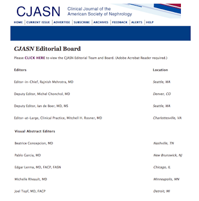
Today I'm part of the visual abstract editorial team for CJASN.

I'm proud that nephrology, represented by CJASN, AJKD and JASN, has moved swiftly to adopt this new medium.

The team is working collaboratively and each of us is signing off on all of the designs so we can continue with #DesignThinking without breaking confidentiality. I'm really proud of the work we have done so far:


Related Posts Plugin for WordPress, Blogger...